Competition for Particle Detection in 3D Cryo-Electron Tomography
Develop a ML model for annotating subcellular structures and proteins in CryoET data
$75,000 in prizes
931 Teams / 6855 Entrants
27,971 Submissions
76 Countries
10 Winning Teams
How is this score calculated
1st Place
Score: 0.78759
Daddies
Members: - Christof Henkel,
- Eugene Khvedchenya
- Christof Henkel,
- Eugene Khvedchenya
The 1st place solution employs an ensemble approach combining segmentation models (3D UNets with ResNet & B3 encoders) and object detection models (SegResNet and DynUnet backbones) from MONAI. Segmentation uses weighted CrossEntropy loss (256:1 positive:negative weighting), while detection implements a modified PP-Yolo loss with IoU-based point-point similarity metrics. Models are trained on 96×96×96 patches with inference on larger volumes, and both approaches are merged through a novel scaling technique that aligns feature map distributions before object detection post-processing. Performance is optimized by converting models to TensorRT, achieving a 200% speedup and enabling parallel inference on two T4 GPUs.
2nd Place
Score: 0.78381
LuoZiqian&Lion
Members: - Ziqian Luo,
- Shuo Wang
- Ziqian Luo,
- Shuo Wang
The 2nd place solution employs an ensemble of multiple lightweight segmentation models with parameter sizes ranging from 873K to 14.2M, including architectures such as UNet3D, VoxResNet, VoxHRNet, SegResNet, DenseVNet, and UNet2E3D. The models are trained using Tversky Loss, Dice Loss, and Cross-Entropy Loss with customized mask radii for each particle type, utilizing InstanceNorm3d and PReLU for enhanced training stability. After segmentation, particle centroids are computed using CC3D and filtered based on voxel count statistics, with an ensemble strategy involving averaging of 7 to 10 complementary models and test-time augmentation.
3rd Place
Score: 0.78351
ONCE UPON A MOON
Members: - tangtang1999
- tangtang1999
The 3rd place solution implements an ensemble of 3D UNet models with ResNet101 backbone, trained using Cross Entropy loss on all seven particle classes including the non-scored beta-amylase. Training utilizes smaller input dimensions (64×128×128) while inference benefits from larger dimensions (64×256×256), coupled with Exponential Moving Average (EMA) with a decay of 0.995 for model stabilization. The final submission consists of a 4-fold average ensemble (from 7-fold cross-validation) with test-time augmentation including flips along x, y, z axes and 90-degree rotations in the x-y plane.
Competition Details
About the Competition
We held a competition for the development of machine learning algorithms to overcome a major bottleneck limiting biomedical discoveries—the annotation and analysis of high resolution 3D images from advanced imaging technologies.
Goal
Advance the understanding of cell biology through machine learning algorithms to annotate particles in 3D images of cells captured by cryoET.
The resulting algorithms were able to perform robust annotation of particles of variable shapes and sizes within the hundreds of 3D images in the competition dataset after being trained on a limited set of available reference annotations from the same dataset.
Competition Data
Competition Deposition Name:
CZII - 2024 CryoET Object Identification Challenge
Experimental and simulated training data for the CryoET Object Identification Challenge. Each dataset contains tilt series, alignments, tomograms and ground truth annotations for six protein complexes (Apo-ferritin, Beta-amylase, Beta-galactosidase, cytosolic ribosome (80S), thyroglobulin and VLP). Curation procedures are described in detail in the accompanying paper. Details on how the dataset was used in the competition are available on Kaggle.

Challenge Resources
To reduce the onboarding time for competitors, an extensive set of example notebooks was provided. These notebooks include the following models:
These notebooks also leverage the copick library for handling cryoET datasets, for which we provided PyTorch Datasets and utility functions to simplify the creation of data loaders, metadata tracking, and model performance analysis.
These example notebooks can be found in the Github Repository - CZII ML Challenge Notebooks.
In addition, the CryoET Data Portal provides multiple annotated datasets of protein complexes in situ which can be used to tune machine learning algorithms to the crowded nature of in situ samples.
What is CryoET?
Overview
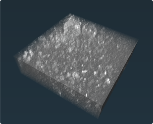
Cryo-electron tomography (CryoET) is an imaging technique that enables 3D visualization of the cell at sub-nanometer resolution but, unlike other high-resolution imaging techniques, the cryogenic (frozen) condition preserves cellular architecture so this detailed view includes protein structures in their natural biological context. Three-dimensional tomograms can be generated from many images of a thin slice through a cell, taken while tilting the specimen in multiple directions.
A given tomogram is typically only about 200 nanometers thick—approximately five hundred times thinner than a sheet of paper—yet packed with information about the structures of the cellular machinery driving health and disease.
For more information about CryoET basics, check out the educational articles from the CryoET Data Portal documentation site.
Glossary of Terms
80S ribosome
- Ribosomes are the molecular machines that translate the genetic information from the intermediary mRNA templates into proteins. The 80S ribosome is specifically the eukaryotic ribosome, and it is abundant in cell lysate since translation of mRNA is a constant activity within the cell.
Apo-ferritin
- A 24-subunit globular protein in all cells and tissues. It binds and transports iron in all cells and tissues. Apoferritin refers to the iron-free form of the protein.
Annotating
- The process of identifying proteins or membranes of interest in noisy 3D tomograms.
- See also labeling and picking.
Competition Contributors
Contributors are listed alphabetically by last name:
- David Agard
- Richa Agarwal
- Ashley Anderson
- Jeremy Asuncion
- Rodrigo Baltazar
- Tristan Bepler
- Nina Borja
- Alister Burt
- Bridget Carragher
- Mikala Caton
- Anchi Cheng
- Chi-Li Chiu
- Yongbaek Cho
- Ellaine Chou
- Bryan Chu
- Charlie Dubbledam
- Kira Evans
- Kirsty Ewing
- Jessica Gadling
- Lorenzo Gaifas
- Kyle Harrington
- Matthias Haury
- Utz Heinrich Ermel
- Norbert Hill
- Erin Hoops
- Timmy Huang
- Peng Jin
- Ann E Jones
- Saugat Kandel
- Kandarp Khandwala
- Robert Kiewisz
- Dari Kimanius
- Mykhailo Kopylov
- Justine Larsen
- Manuel Leonetti
- Donghui Li
- Emma Lundberg
- Kristen Maitland
- Gorica Margulis
- Dannielle McCarthy
- Elizabeth Montabana
- Ben Nelson
- Jun Xi Ni
- Stephani Otte
- Mohammadreza Paraan
- Noeli Pazsoldan
- Ariana Peck
- Clinton Potter
- Janeece Pourroy
- Dana Sadgat
- Simon Sander
- Jonathan Schwartz
- Daniel Serwas
- Shu-Hsien Sheu
- Hannah Siems
- Trent Smith
- Andrew Sweet
- Shivanshi Vaid
- Madhuri Vangipuram
- Manasa Venkatakrishnan
- Carmela Villegas
- Thorsten Wagner
- Eric Wang
- Zun Shi Wang
- Feng Wang
- Samantha Yammine
- Yue Yu
- Zhuowen Zhao
- Shawn Zheng
- Ellen Zhong
Sponsored By:


