Deposition
Cryo-electron tomography of axons and varicosities in cultured primary mouse dorsal root ganglion neurons (baseline data and annotations)
- Deposition ID:CZCDP-10362
Release Date: 2026-08-14
Last Modified: 2026-08-14
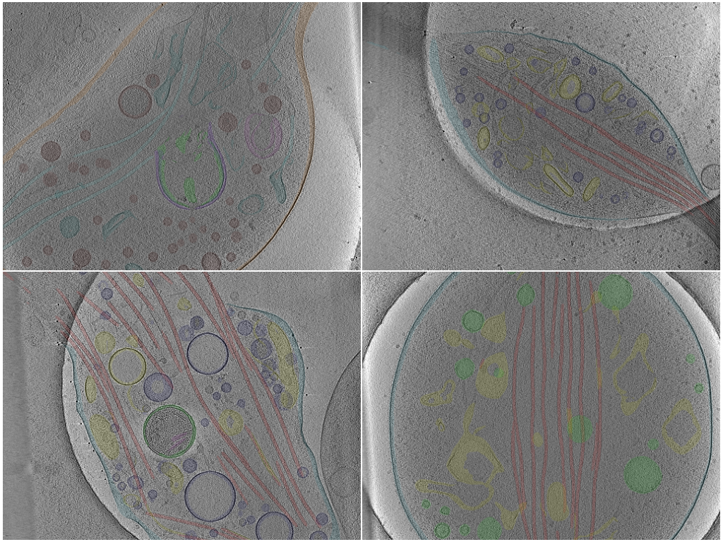
Photo Caption: Central slab of 4 representative tomograms with selected sets of annotations.
Deposition Overview
Cryo-electron tomography of intact, unmilled peripheral axons and their varicosities in primary dorsal root ganglion neurons dissociated from 3-month-old mice and cultured for 48 h on gold Quantifoil grids. Neurons were plunge-frozen without fixation or staining, and the thin axonal processes growing over the grid were imaged directly. Most tilt series were collected on a Titan Krios at 300 kV with a Gatan energy filter and a K2 Summit detector; a comparative subset was collected on a Glacios at 200 kV with a Selectris energy filter and a Falcon 4i detector. No differences in structural information were noted between the two instruments and the data were pooled for analysis. Tomograms were reconstructed by weighted back-projection in IMOD and denoised by IsoNet deconvolution. Organelle membranes were pre-segmented with MemBrain-seg and microtubules with TARDIS-MT, then manually curated in Amira into a multi-class semantic scheme spanning the plasma membrane, microtubules, mitochondria, endoplasmic reticulum and vesicles enclosed within it, multivesicular bodies, multilamellar vesicles, endosomes, phagophores and free cytoplasmic vesicles. The deposition holds 105 tomograms, drawn from the 192 reconstructed for the study, together with their raw movies and tilt series; all of them carry a segmentation.
Deposition Data
Annotations:776
Tomograms:134
Publications
Related Databases
Not Submitted
Methods Summary
Annotations | Method Type | Method Details | Method Links |
|---|---|---|---|
| 776 | Hybrid | MemBrain-seg (membranes) and TARDIS-MT (microtubules) pre-segmentation, manually curated into organelle classes in Amira on 4x-binned, IsoNet-deconvolved tomograms. | Source Code:IsoNet source code +17 more |
Deposited Data
Data Contents
Filter by:
776 of 776 Annotations
Annotation Name | Object Shape Type | Method Type | Deposited In |
|---|---|---|---|
109endoplasmic reticulum Annotation ID: AN-182956 | SegmentationMask | Hybrid | |
108cytoplasmic vesicle Annotation ID: AN-182955 | SegmentationMask | Hybrid | |
103microtubule Annotation ID: AN-182954 | SegmentationMask | Hybrid | |
102plasma membrane Annotation ID: AN-182953 | SegmentationMask | Hybrid | |
101mitochondrial inner membrane Annotation ID: AN-182952 | SegmentationMask | Hybrid | |
100mitochondrial outer membrane Annotation ID: AN-182951 | SegmentationMask | Hybrid | |
110endoplasmic reticulum Annotation ID: AN-182950 | SegmentationMask | Hybrid | |
108cytoplasmic vesicle Annotation ID: AN-182949 | SegmentationMask | Hybrid | |
103microtubule Annotation ID: AN-182948 | SegmentationMask | Hybrid | |
102plasma membrane Annotation ID: AN-182947 | SegmentationMask | Hybrid | |
100mitochondrial outer membrane Annotation ID: AN-182946 | SegmentationMask | Hybrid | |
114endoplasmic reticulum Annotation ID: AN-182945 | SegmentationMask | Hybrid | |
112endosomal intralumenal vesicle Annotation ID: AN-182944 | SegmentationMask | Hybrid | |
111endosome Annotation ID: AN-182943 | SegmentationMask | Hybrid | |
108cytoplasmic vesicle Annotation ID: AN-182942 | SegmentationMask | Hybrid | |
105vesicle Annotation ID: AN-182941 | SegmentationMask | Hybrid | |
102plasma membrane Annotation ID: AN-182940 | SegmentationMask | Hybrid | |
101mitochondrial inner membrane Annotation ID: AN-182939 | SegmentationMask | Hybrid | |
100mitochondrial outer membrane Annotation ID: AN-182938 | SegmentationMask | Hybrid | |
111endoplasmic reticulum Annotation ID: AN-182937 | SegmentationMask | Hybrid |
- 1
- 2
- 3
- 4
- 5
- 39