Deposition
A generalizable and targeted molecular biopsy approach for in situ cryogenic electron tomography of vitreous brain tissue
- Deposition ID:CZCDP-10357
Release Date: 2026-07-09
Last Modified: 2026-07-09
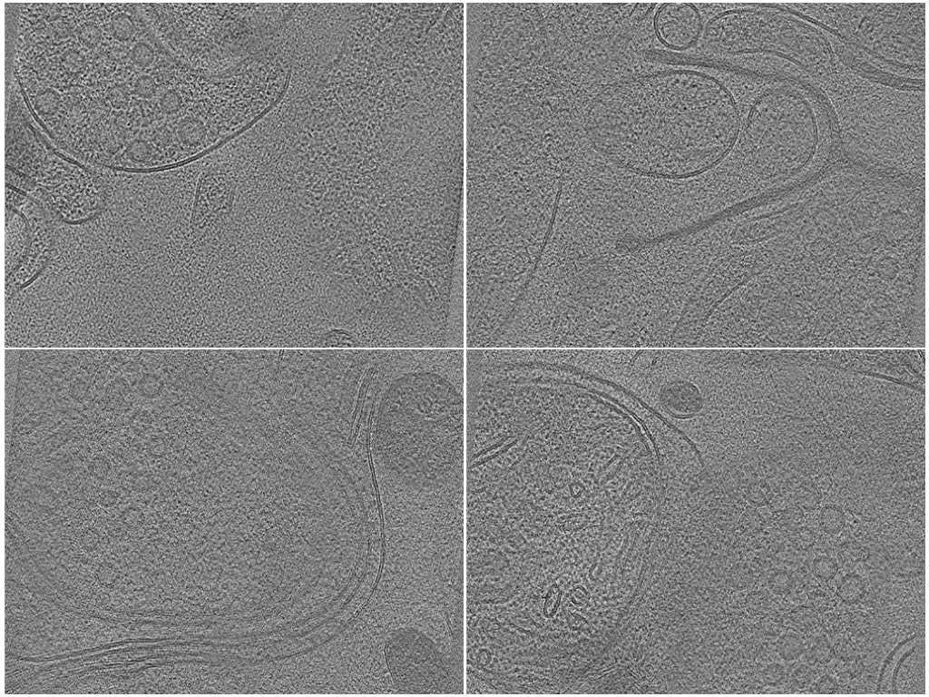
Photo Caption: Central slab of 4 representative tomograms with selected sets of annotations.
Deposition Overview
This deposition contains in situ cryo-electron tomography (cryo-ET) data of vitreous mouse brain tissue produced by a targeted molecular-biopsy workflow. Adult mouse hippocampal sections were preserved by high-pressure freezing, screened and targeted by cryo-fluorescence microscopy, and thinned to electron-transparent lamellae by xenon/argon plasma focused ion beam (PFIB) milling using perpendicular and planar cryo-lift-out geometries. This approach targeted sub-regions of the hippocampal CA1 field, spanning stratum pyramidale (CA1-sp) through stratum radiatum (CA1-sr) toward stratum lacunosum-moleculare, as well as the cerebral cortex, across biopsies from several adult mice.
Authors
Methods Summary
Annotations | Method Type | Method Details | Method Links |
|---|---|---|---|
| 423 | Automated | Automated membrane segmentation of the reconstructed tomograms using membrain-seg, a deep-learning membrane segmentation model, restricted to the valid-sample volume: the valid-sample mesh was filled into a mask (copick convert mesh2seg) and only membrane voxels inside it were kept (copick logical clipseg). The output is a single binary membrane mask per tomogram, with no manual correction step. | Source Code:copick +7 more |
| 423 | Automated | Boundary of the valid specimen volume within each tomogram. A 3D U-Net (octopi) predicts the sample region; the prediction is cleaned to its largest connected component and a slab mesh with smooth parallel top and bottom surfaces is fitted to it, then intersected with the tomogram's valid reconstruction area to yield a closed mesh enclosing the usable specimen volume. | Source Code:copick +6 more |
Deposited Data
Data Contents
Filter by:
846 of 846 Annotations
Annotation Name | Object Shape Type | Method Type | Deposited In |
|---|---|---|---|
101sample Annotation ID: AN-183802 | Mesh | Automated | |
100membrane Annotation ID: AN-183801 | SegmentationMask | Automated | |
101sample Annotation ID: AN-183800 | Mesh | Automated | |
100membrane Annotation ID: AN-183799 | SegmentationMask | Automated | |
101sample Annotation ID: AN-183798 | Mesh | Automated | |
100membrane Annotation ID: AN-183797 | SegmentationMask | Automated | |
101sample Annotation ID: AN-183796 | Mesh | Automated | |
100membrane Annotation ID: AN-183795 | SegmentationMask | Automated | |
101sample Annotation ID: AN-183794 | Mesh | Automated | |
100membrane Annotation ID: AN-183793 | SegmentationMask | Automated | |
101sample Annotation ID: AN-183792 | Mesh | Automated | |
100membrane Annotation ID: AN-183791 | SegmentationMask | Automated | |
101sample Annotation ID: AN-183790 | Mesh | Automated | |
100membrane Annotation ID: AN-183789 | SegmentationMask | Automated | |
101sample Annotation ID: AN-183788 | Mesh | Automated | |
100membrane Annotation ID: AN-183787 | SegmentationMask | Automated | |
101sample Annotation ID: AN-183786 | Mesh | Automated | |
100membrane Annotation ID: AN-183785 | SegmentationMask | Automated | |
101sample Annotation ID: AN-183784 | Mesh | Automated | |
100membrane Annotation ID: AN-183783 | SegmentationMask | Automated |
- 1
- 2
- 3
- 4
- 5
- 43