Dataset
Cryo-electron tomography of centrosomes in C. elegans embryonic cells
- Dataset ID:DS-10477
Release Date: 2026-03-26
Last Modified: 2026-03-26
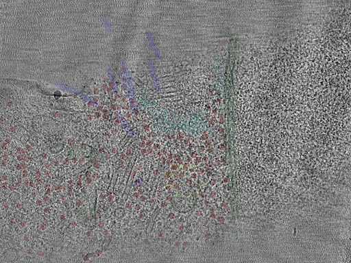
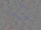
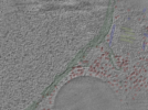
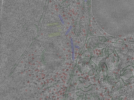
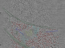
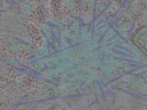
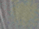
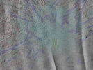
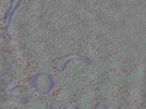
Photo Caption: Central slab from a representative tomogram of a Caenorhabditis elegans sample with selected set of annotations.
Dataset Overview
Cryo-electron tomography data of centrosomes in dissociated C. elegans embryonic cells across the cell cycle. Centrosomes were visualized in cells isolated from early-stage embryos (strain JWW69 expressing GFP::SPD-5 and H2B::mCherry). Cells were plunge-frozen, thinned by cryo-FIB milling, and imaged on a 300 kV Titan Krios with a K2 Summit detector, Volta phase plate, and energy filter. 12 tomograms were collected at centrosomes in interphase, prophase, metaphase, anaphase, and telophase stages. Comprehensive annotations of centrioles, microtubules, ribosomes, membranes, and pericentriolar material are provided. Tilt series were aligned using patch tracking in IMOD, and tomograms were reconstructed using Warp.
Publications
Related Databases
EMPIAR ID:EMPIAR-12049
Runs
12 of 12 Runs
Run Name | Tilt Series Quality Score | Objects | ||
|---|---|---|---|---|
 | cent1 Run ID: RN-33577 | 2 - Poor |
| |
 | cent10 Run ID: RN-33578 | 5 - Excellent |
| |
 | cent11 Run ID: RN-33579 | 3 - Moderate |
| |
 | cent12 Run ID: RN-33580 | 5 - Excellent |
| |
 | cent2 Run ID: RN-33581 | 5 - Excellent |
| |
 | cent3 Run ID: RN-33582 | 5 - Excellent |
| |
 | cent4 Run ID: RN-33583 | 5 - Excellent |
| |
 | cent5 Run ID: RN-33584 | 5 - Excellent |
| |
 | cent6 Run ID: RN-33585 | 5 - Excellent |
| |
 | cent7 Run ID: RN-33586 | 4 - Good |
| |
 | cent8 Run ID: RN-33587 | 3 - Moderate |
| |
 | cent9 Run ID: RN-33588 | 3 - Moderate |
|