Dataset
Cryo-electron tomography of axons and varicosities in cultured primary mouse dorsal root ganglion neurons (Titan Krios, 30-31 July 2025)
- Dataset ID:DS-10512
Release Date: 2026-08-14
Last Modified: 2026-08-14
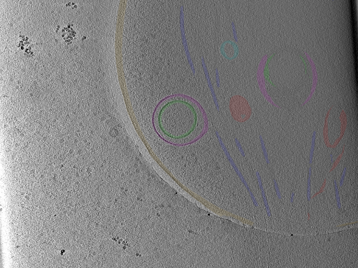
Photo Caption: Central slab from a representative tomogram of a Mus musculus sample with selected set of annotations.
Dataset Overview
Cryo-electron tomography of intact, unmilled peripheral axons and their varicosities in primary dorsal root ganglion neurons dissociated from 3-month-old mice and cultured for 48 h on gold Quantifoil grids. The axons grow thin enough over the grid surface to be imaged directly, with no cryo-sectioning and no focused-ion-beam milling, so the varicosities (roughly 0.6-1.9 microns across) and the much narrower shafts connecting them are captured in a near-native state. Tilt series were collected from -51 to +51 degrees in 3 degree increments on a Titan Krios operated at 300 kV, equipped with a Gatan energy filter and a K2 Summit direct electron detector. Tomograms were reconstructed by weighted back-projection in IMOD and denoised by IsoNet deconvolution. This dataset holds 31 tomograms, all of which carry a manually curated multi-class organelle segmentation covering the plasma membrane, microtubules, mitochondrial outer and inner membranes, endoplasmic reticulum and vesicles enclosed within it, multivesicular bodies, multilamellar vesicles, endosomes, phagophores and free cytoplasmic vesicles. The study uses these to ask how the spatial confinement of an axon constrains organelle architecture.
Publications
Related Databases
Not Submitted
Runs
5 of 32 Runs
Run Name | Tilt Series Quality Score | Objects | ||
|---|---|---|---|---|
 | Position_2 Run ID: RN-36227 | 5 - Excellent |
| |
 | Position_20 Run ID: RN-36228 | 5 - Excellent |
| |
 | Position_30 Run ID: RN-36237 | 5 - Excellent |
| |
 | Position_32 Run ID: RN-36238 | 5 - Excellent |
| |
 | Position_42 Run ID: RN-36246 | 5 - Excellent |
|